As Doctoral Candidate DC09 in the EU-funded
HER-CARE MSCA Doctoral Network
, I investigate who may benefit from supplemental imaging and when longer screening intervals may be appropriate. The planned work includes external validation across Swedish and US screening populations.
Research direction · clinical benefit to be evaluated
Doctoral workflow, imaging examples & credits +
THE RESEARCH PIPELINE
From breast imaging to future risk.
Illustrative imaging examples and a conceptual modelling workflow.
01 / IMAGING & CONTEXT
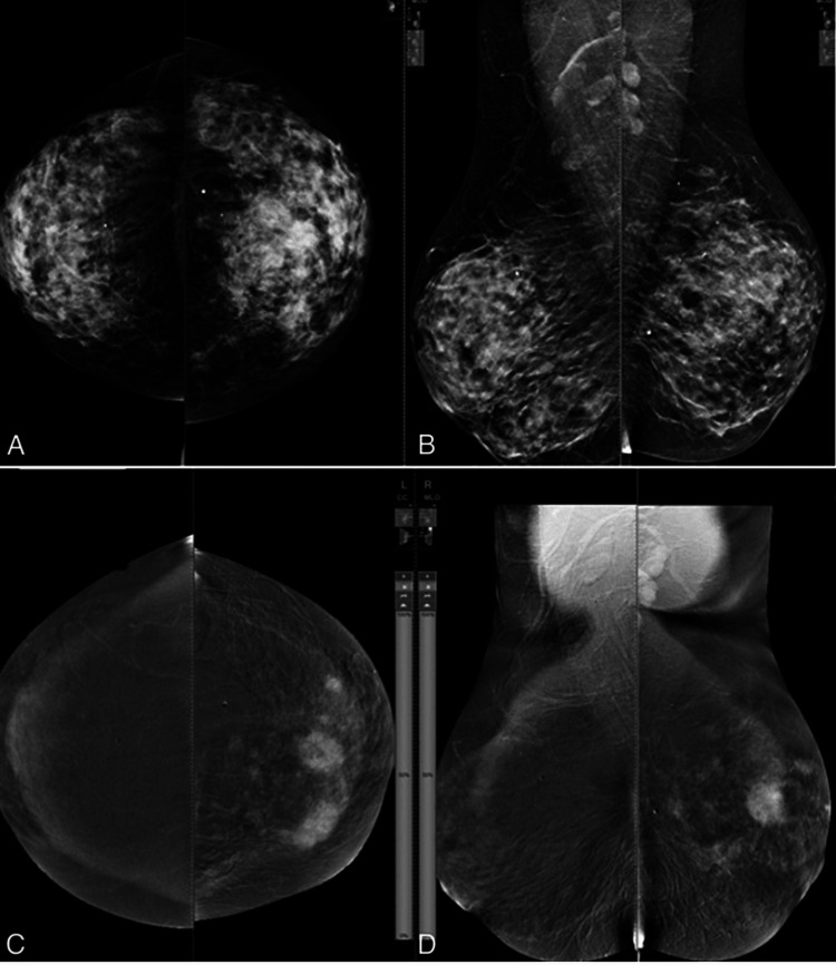
Mammography
Standard & contrast-enhanced views
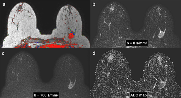
Breast MRI
Complementary imaging information
Age & family history
Breast density
Clinical & genetic risk factors
Single examinations and longitudinal mammograms, where available.
02 / LEARN & INTEGRATE
Deep learning & multimodal modelling
Extract image representations and combine them with clinical risk factors.
Image-derived features
Clinical feature integration
Time-to-event risk modelling
Evaluate discrimination, calibration, and performance across populations.
03 / PREDICT & EVALUATE
Short-term breast cancer risk
Baseline
1 year
3 years
5 years
Higher risk
Who may benefit from supplemental imaging?
Lower risk
When might longer screening intervals be appropriate?
After a negative MRI
How does subsequent risk change over time?
Research aims, not individual predictions or clinical recommendations.
Conceptual overview of planned research; no model outputs or study results are shown. MRI and mammography examples depict published cancer cases, not negative screening examinations or participants in my PhD cohorts. Modalities illustrate complementary research streams; MRI is not assumed to be available for every participant.
Image sources & credits
+
Breast MRI: Camps-Herrero J. (2019),
Diffusion-weighted imaging of the breast
, reproduced as Figure 4 in the same review.
CC BY 4.0
; figure reproduced unchanged. Neural network and risk timeline are explanatory schematics created for this site.